 The historical development of medical thermography
Early infrared imaging systems were developed during the 1940s and became available to industry and medicine first in 1959.
The Pyroscan
(a 1942 instrument) was first used in Bath in 1959 and was used to image the increased heat over arthritic joints. Picture quality
improved with the Mark 2 instrument, although each image took 34 minutes to acquire and was almost impossible to quantify. Later, with
improved equipment, better dynamic (and objective images) were obtained that could usefully supplement radiological investigation.
During the 1960s and 1970s a new generation of thermal imaging systems were developed in Europe, the USA and Japan. Oscilloscope displays
were introduced and electronic isotherms were added to the image. By multiple exposure colour photography, the first colour thermograms were
produced in the 1960s. Mini-computers for image processing arrived in the mid- to late 1970s providing colour displays, image analysis and,
importantly, data and image storage.
This marked the beginning of quantitative thermography.
Modern systems introduced within the last three years use focal plane array detectors, with high speed images at high thermal and spatial
resolution.
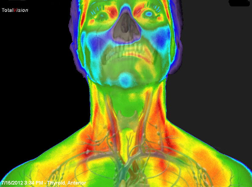
Image quality has dramatically improved, modern digital thermograms are now very different from the crude clinical images obtained
since 40 years ago.
Technological advances in infrared cameras within the last few years have promoted MIT as a powerful measurement tool. A new generation of
high-resolution cameras, appropriate software and standardized protocols have been developed for medical imaging, resulting in improved
diagnostic capability and reliability. In 1987, the American Medical Association recognized MIT as a feasible diagnostic tool.
|